Alkaline Lysis Miniprep of Plasmid DNA from E.coli
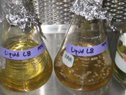
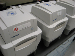
For Alkaline lysis a single bacterial colony was inoculated in 3ml of LB containing appropriate antibiotics, and grown overnight at 37°C. 1.5ml of the culture was transferred to an Eppendorf tube and the bacterial cells were pelleted by centrifugation for two minutes at 14,000 rpm in a microcentrifuge (Eppendorf).
 The pellet was resuspended in 500 ml of STE (100mM NaCl, 10mM Tris.Cl pH 8.0, 1mM EDTA) and re-pelleted by centrifugation. The pellet was resuspended in 100 m l ice-cold Solution I (25mM Tris.Cl, 50mM EDTA, pH8.0, 4mg/ml lysozyme) by vortexing. 200 m l of freshly prepared solution II (0.2M NaOH, 1% SDS) was added, and mixed by slowly inverting the tube five times. Following a 5 minute incubation at room temperature 150 m l of ice-cold solution III (3M/5M KOAc, pH 4.8) was added and mixed by vortex. Samples were incubated on ice for five minutes, then centrifuged 14000 rpm (Eppendorf microcentrifuge) to remove cell debris. The supernatant was transferred to an Eppendorf tube, and DNA was ethanol precipitated. The DNA pellet was dissolved in 50 m l TE pH 8.0 or dH 20.
Phenol/Chloroform Extraction
To remove contaminating proteins, Phenol/chloroform solution (24 Phenol: 24 Chloroform: 1 Isoamyl alcohol (equilibrated with Tris.Cl pH 8.0; Biogene ltd.)) was added at an equal volume to the DNA samples. Samples were mixed by vortexing, then centrifuged to separate different phases. The aqueous, DNA containing phase was transferred to a fresh Eppendorf tube and DNA precipitated.
DNA Precipitation
DNA was precipitated in either ethanol or isopropanol. For isopropanol precipitation 0.7 volumes of isopropanol was added to the DNA sample, and incubated at room temperature for 20 minutes. For ethanol precipitation, one-tenth volume 3M NaOAc (pH 5.2) and 2 volumes of 100% ethanol were added to the DNA sample. Followed by a 20 minute incubation at –20°C. In each case, DNA was pelleted by centrifugation at 14,000 rpm in a microcentrifuge (Eppendorf). The pellet was washed in 70% ethanol, re-pelleted and centrifuged.
Genomic DNA Extraction
Small-scale extraction of genomic DNA from Arabidopsis leaves was achieved by using a variation of the Dellaporta protocol (Dellaporta et al., 1983). Frozen leaf tissue is first ground to a fine powder in an Eppendorf. To this is added 750ml of extraction buffer (100mM Tris pH 8.0, 50mM EDTA pH8.0, 500mM NaCl, 10mM b -mercaptoethanol); and 50 m l of 10% SDS (to dissolve the cell membranes).
After incubation at 65°C, 250 m l of 5M potassium acetate is added. This is incubated on ice for 20 min; the solution is then separated by centrifugation (14000 rpm for 10 min) (Eppendorf microcentrifuge). 500 ml of isopropanol is added to the supernatant, which is incubated for 20 min at –20 °C. After further centrifugation (14000 rpm for 10 min) (Eppendorf microcentrifuge), the supernatant is discarded and the genomic DNA pellet dried in a drying centrifuge for 5 seconds. The pellet is re-dissolved in 100 m l of 10mM TrisHCL or DH 2O and stored at -20 oC until required.
Agrobacterium and Bacterial Cell Transformation
Bacterial Cell Transformation
50 ml of subcloning efficiency DH5a TM competent cells (2.5 x 10 8 cfu/mg pUC19) (Life Technologies) are gently thawed on ice in an Eppendorf tube. 1-10ng of plasmid DNA is added to the tube, and mixed by flicking the tube. The sample is then incubated on ice for 30 minutes. The cells are heat shocked at 42°C for 45 seconds and chilled on ice for two minutes. 950 ml of LB broth was added to the cells, and they are incubated for one hour at 37°C. Cells were then plated onto LB-agar with the appropriate antibiotics.
Agrobacterium cell transformation
Competent Agrobacterium cells ( C58C1 pGV101 pMP90) are transformed by electroporation. Firstly 40 ml of cells should be thawed on ice. The binary vector plasmid and a helper plasmid pSOUP, added to the cells, and mixed by flicking, and transferred to a pre-cooled electroporation cuvette (Biorad). An electric pulse is applied across the cuvette (field strength = 2.5kV/cm, Capacitance = 25 m F, resistance = 400 W , pulse-length = 8-12mS). The cells are transferred to 1ml YT broth and incubated at 30°C for 3 Hrs. The sample is centrifuged and the top 900ml of broth is discarded. The pelleted cells are resuspended in the remaining 100 ml of medium, plated onto selective YT medium and incubated for 3 days at 30°C.
Running Agarose Gels
Separation of DNA and PCR products was performed by electrophoresis using agarose gels. The density of gel used depends upon product length. DNA and PCR samples are prepared for loading by the addition of 10X loading dye (30% glycerol, 0.25% Xylene Cyanol, 0.25% Bromophenol Blue) and dH 2O.
Constructing Agarose Gels
To make up the agarose gels for electrophoresis, agarose and 0.5X TBE are mixed to the relevant concentration (typically 0.8 to 4% w/v). The solution is heated in a microwave oven until boiling. The solution is allowed to cool to approximately 65 oC and ethidium bromide added. The solution is poured into a gel holder and allowed to set. At this stage it is placed into a gel tank (Bio-Rad) containing 0.5X TBE buffer. Following loading, the samples are electrophoresed at between 50V and 125V on the agarose gels. Following electrophoreses DNA and PCR products are visualised in UV light by use of a UV transilluminator, and the image captured using the Gel DOC 2000 TM (BIO-RAD) system.
Polymerase Chain Reaction - PCR
PCR (Polymerase chain reaction) are typically carried out in 25 ml reactions. For standard PCRs, such as used for mapping, DNA is amplified by use of Taq DNA polymerase (Roche).
 If a specific product is required, such as was used when cloning, then a proof reading polymerase such as pwo or Expand-HiFidelity (Roche) should be used. Typically a PCR mixture constituted 1.25 Units Polymerase, 0.4 mM Fwd and Rev primers, 200 mM dNTPs (dATP, dCTP, dGTP, dTTP), and 1xPCR buffer (10x PCR buffer: 100mM Tris.Cl pH 8.3, 500mM KCl, 15mM MgCl 2, 0.1% gelatin, 0.5% tween ® 20, 250 m g/ml BSA).
Polymerase chain reaction (PCR) amplification consists of an initial two minute denaturing step at 96°C. Ffollowed by a cycle of denaturing, annealing and extension conditions. The number of cycles and the temperatures of each step is variable. A typical PCR has 30 cycles of (i) 30 secs denaturing at 94°C (ii) 30 secs annealing at 62°C (iii) 2 minute extension at 72°C. This cycling is followed by a 10 minute final extension. The main changes in PCR conditions involves varying the number of cycles performed and the use of different annealing temperatures.